
Giemsa staining: An overview, principle, and applications
Introduction
Malaria was rampant worldwide even towards the mid of the 20th century. Even though it was established that malaria was caused by a parasite and can be diagnosed by observing blood smears, a validated technique for staining and observing the blood smears on a large scale was unavailable. In 1902, Gustav Giemsa formulated the Giemsa stain to observe blood smears more reliably. Other researchers further modified it, which led to many variations.
Giemsa staining was so efficient that it is used for blood smears and acts as a gold standard in malarial testing. Though the technique was devised for malaria, it is now widely used in other fields such as hematology, cytology, histology, bacteriology, and off-late in cytogenetics.
Principle
Gustav Giemsa formulated the stain to be a mix of a cationic dye Azure II (the major component is Azure B), and an anionic dye – eosin Y. Azure B is a variant of methylene blue after its oxidation. Since these dyes have different charges, they have an affinity toward different molecules/cellular compartments based on their charge. For example, Azure B being basic, react with acidic components like nucleic acid, specifically to the phosphate groups and in AT-rich regions of DNA, producing a deep purple color (stains the whole of the nucleus purple). While Eosin is acidic in nature and reacts with the cytoplasm and cytoplasm components to give a pink color.
Applications
Parasitology
Blood smears (both thin and thick) are observed to diagnose malaria, or Trypanosoma (Fig 1) can be observed to conclude the presence of infection. Parasites are stained purple because of nucleic acids, while the RBC are either colorless or stained a faint pink. The staining technique is also used for Histoplasma, Leishmania, Toxoplasma, and Pneumocystis.
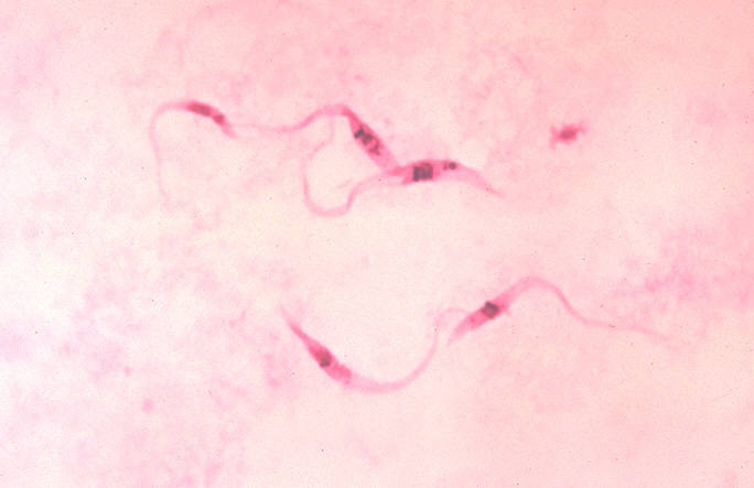
Figure 1. Trypanosoma stained with Giemsa.
Cytogenetics
One of the well-known applications of Giemsa in cytogenetics is karyotyping of the chromosomes. The stian is used for the G banding (Giemsa banding) and R banding (Reverse Giemsa banding). In G staining, chromosomes show purple bands at the AT-rich sequence of DNA. This coloration is due to the fast migration of Azure B (positive charge) into the minor grooves of DNA, which gives a blue color to the DNA. The binding of Azure B also changes the confirmation of DNA such that it accommodates large, negatively charged, slow diffusing eosin Y (red/pink). In DNA, Azure B and eosin Y form a thiazine-eosin precipitate in a 2:1 ratio, respectively. A hydrophobic environment favors precipitation.
Since AT-rich sequences are usually transcriptionally inactive and harbours hydrophobic proteins, the complex formation is favored over here compared to other regions of chromosomes. The applications include but are not limited to diagnosing chromosomal abnormalities (trisomy, transversions, and translocations).
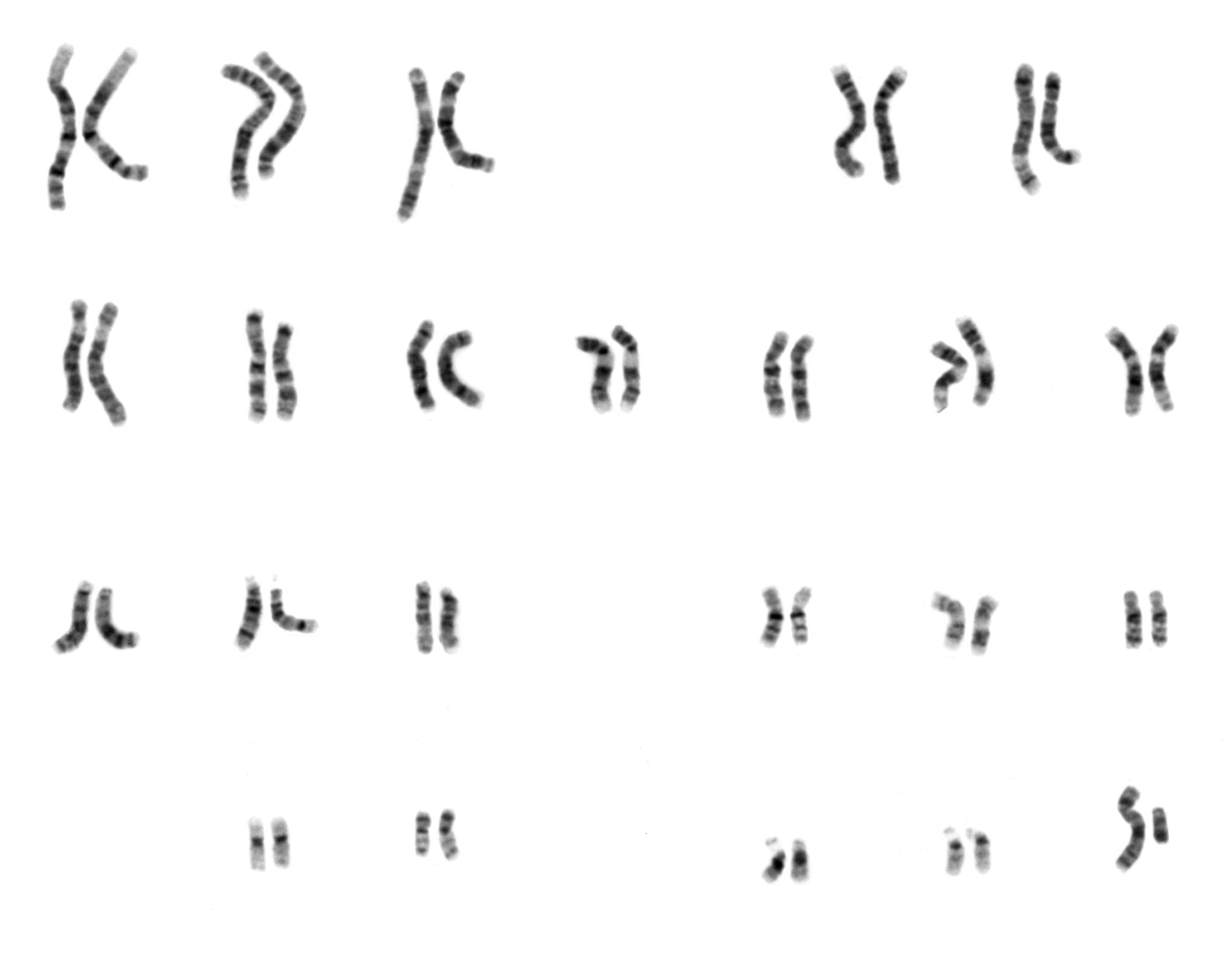
Figure 2. G-banding of human male using Giemsa staining.
Hematology
Thin blood smears can also be obtained for hematological purposes. Each type of WBCs stains differently and can be distinguished morphologically. The characteristics of each cell type after staining would be :
- Eosinophils – purple-blue nucleus, pale pink cytoplasm with red cytoplasmic granules.
- Basophils – purple nucleus and bluish granules.
- Neutrophils – purple-red nucleus and pale pink cytoplasm.
- Lymphocytes – navy blue nucleus and faintly stained blue cytoplasm.
- Monocytes – purple.
- Platelets – small, dark blue without much evident cytoplasm.
Giemsa can also be used to stain bone marrow biopsies to ascertain any pathologies or disease conditions.
Histology/cytology
Giemsa stain can act as an alternative to Hematoxylin-Eosin staining in most histological examinations and carries almost a similar protocol. An underlying pathological condition can be diagnosed by comparing a healthy tissue with a diseased one. For example, in some tumors and cancers, diseased tissues show a higher number of immune cells or WBCs. This can be clearly observed and ascertained. The staining method is known to give sharp details of the endocrine pancreas, the male gonads and bulbourethral glands (staining the cytoplasmic inclusions), and the fibrils of eccrine and sebaceous glands.
Bacteriology
A number of bacteria can be detected by Giemsa staining the tissue they have infected. Examples include staining for stomach biopsies to find Helicobacter pylori infection ( Helicobacter cells appear dark purple compared to pink tissue). Another example would be Yersinia pestis, which can be seen in liver, lung, and kidney biopsies as tiny dark structures against cells that are mostly stained pink or pale blue.
Reference
- Giemsa: The Universal Diagnostic Stain.
- Detection of infection or infectious agents by use of cytologic and histologic stains.
- Histopathological Observation of Immunized Rhesus Macaques with Plague Vaccines after Subcutaneous Infection of Yersinia pestis.
- A Review of the Different Staining Techniques for Human Metaphase Chromosomes.
- The Giemsa Stain: Its History and Applications.
- The composition of stains produced by the oxidation of Methylene Blue.
- Giemsa Stain- Principle, Procedure, Results, Interpretation.
- Blood Cells, A Practical Guide.